The development of algorithms and tools for multidimensional visualization of medical imaging data from different imaging systems. This mainly involves the reconstruction of models, based on the intensity of some or all data points of the original or processed data. Reconstructed multidimensional models are rendered fully (volume rendering) or partially (surface rendering), using a variety of techniques often associated with the gaming industry, such as ray-tracing, ray-casting and anti-aliasing.
Three-dimensional imaging of cancer tissue risk areas in the ovarian region
The evaluation was based on Apparent Diffusion Coefficient (ADC) data values and for the imaging the localized tumor has been superimposed on the patient’s original MRI data. The less risky areas of the tumor are depicted in blue, potentially risky areas of the tumor are depicted in red and areas of moderate risk are depicted in yellow.
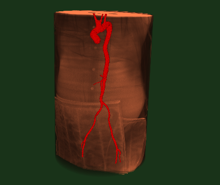
Automatic 3D segmentation of the Aortic Vessel Tree (AVT) in CT images (chest scan)
A CT scan is a diagnostic imaging exam that uses X-ray technology to produce images of the inside of the body. A CT scan can show detailed images of any part of the body, including the bones, muscles, organs and blood vessels.

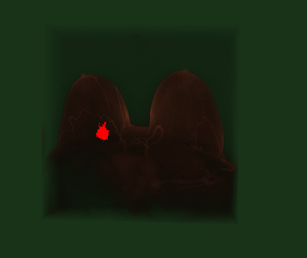
Breast MRI Tumor Automatic Segmentation
MRI scanners create images of the body using a large magnet and radio waves. No ionizing radiation is produced during an MRI exam, unlike X-rays. These images give your physician important information in diagnosing your medical condition and planning a course of treatment.

Non invasive video imaging of retinal blood flow

